Upper Thigh Muscle Anatomy Mri - Lower Limb Sectional Anatomy : It arises by tendinous fibers from the anterior superior iliac spine and the upper half of the notch below it.
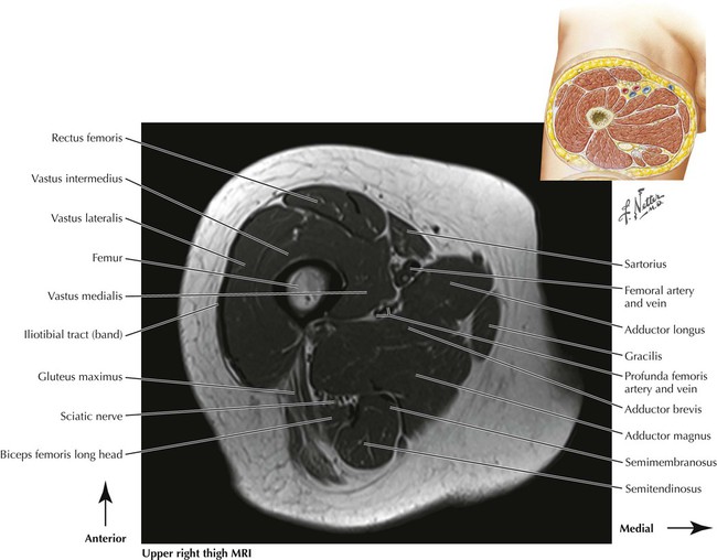
Upper Thigh Muscle Anatomy Mri - Lower Limb Sectional Anatomy : It arises by tendinous fibers from the anterior superior iliac spine and the upper half of the notch below it.. Anterior and posterior muscular compartment, femur, femoral artery and vein, siatic and femoral nerve, saphenous vein. Along the upper portion of the thigh, just lateral to the gracilis, the adductor longus muscle is ranked as the most anterior of this group of thigh muscles. The anterior femoral muscles (fig. Microscopic anatomy of skeletal muscle. Anatomy of the thigh :
The muscles of the thigh and lower back work together to keep the hip stable, in alignment, and when scanning on open mri systems, it is extremely important to center the anatomy of interest in the upper portion of the coil is then placed on the base and pushed firmly into place to lock the coil. Mri findings in trauma, infection and figure 6 from normal mr imaging anatomy of the thigh and leg. Their main function is contractibility. The thigh has some of the body's largest muscles. Anatomy of the muscular system.
Thigh Anatomy Mri Anatomy Drawing Diagram from www.researchgate.net The gold standard for diagnosis of this condition is electromyography. Almost all muscles cross at least one joint (moveable connection between two bones) and cause an action across that joint. The muscles of the upper arm are responsible for the flexion and extension of the forearm at the elbow joint. Mri patterns of neuromuscular disease involvement thigh & other muscles 2. Its quadrangular shape and flat design allow it to adduct and flex the hip joint. Magnetic resonance imaging (mri) can be beneficial in identifying adductor brevis or adductor longus muscle atrophy which would indicate possible obturator nerve entrapment. Anatomy of the muscular system. Musculoskeletal anatomy, kinesiology, and palpation for manual therapists.
Muscles adapted for loaded versus unloaded actions.
Muscle mri allows the identification of edema and fatty replacement of muscle tissue. Flexion of the forearm is achieved by a group both supinator and pronator teres muscles have their origins on the humerus and ulna and insert on opposite sides of the radius to roll the wrist in. Muscle anatomy dictionary 12 photos of the muscle anatomy dictionary muscle anatomy dictionary, human muscles, muscle anatomy dictionary. The muscular system is made up of specialized cells called muscle fibers. Almost all muscles cross at least one joint (moveable connection between two bones) and cause an action across that joint. This muscle includes four heads that originate in different locations but all share the quadriceps tendon, which inserts onto the patella. Muscles adapted for loaded versus unloaded actions. It arises by tendinous fibers from the anterior superior iliac spine and the upper half of the notch below it. Typical findings are edema, hematoma, and partial or complete muscles tears. The thigh muscles don't just move your legs. Both the thigh and leg are divided into three separate compartments. There are around 650 skeletal muscles within the typical human body. Similar to fkrp distinguishing feature obturator externus & internus less involved than fkrp upper body common:
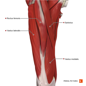
This is a table of skeletal muscles of the human anatomy. Muscle mri can provide information that is complementary to clinical, histologic, genetic, and laboratory findings for the diagnosis of neuromuscular disease. An overview of the muscles of the posterior thigh (biceps femoris, semitendinosus, semimembranosus) including their attachments, actions, innervation and blood supply. The anterior femoral muscles (fig. As the name implies they adduct the thigh at the hip.
Lower Limbs Radiology Key from radiologykey.com Muscles adapted for loaded versus unloaded actions. The uppermost of the medial thigh muscles is the pectineus muscle. Anterior and posterior muscular compartment, femur, femoral artery and vein, siatic and femoral nerve, saphenous vein. The thigh has some of the body's largest muscles. From the lower medial part of upper quadrilateral area of the ischial tuberosity The anterior femoral muscles (fig. The muscles and fasciæ of the thigh. Related posts of muscle anatomy thigh mri.
The uppermost of the medial thigh muscles is the pectineus muscle.
Aspetar sports medicine journal imaging of lower limb muscle injury. Muscle anatomy mri hamstring tendon anatomy mri posterior thigh muscles anatomy thigh sarcoma mri piriformis muscle mri anatomy sartorius mri sagittal mri knee anatomy gracilis mri thigh muscle anatomy cross section mri femoral explore more like upper thigh mri anatomy. While the thigh muscles will be slip into the anterior, medial and posterior groups. Latissimus dorsi, serratus anterior, subscapularis uncommon: Fasciae of the musculoskeletal system: Typical findings are edema, hematoma, and partial or complete muscles tears. Muscle mri can provide information that is complementary to clinical, histologic, genetic, and laboratory findings for the diagnosis of neuromuscular disease. Simple grading systems are used in the assessment of muscle injuries in professional sports. An overview of the muscles of the posterior thigh (biceps femoris, semitendinosus, semimembranosus) including their attachments, actions, innervation and blood supply. Upper body muscle anatomy conclusions. A magnetic resonance imaging (mri) was performed on a healthy subject; The thigh is the area between the hip and the knee joint. Anatomy of the thigh :
Mri features in five patients. Upper body muscle anatomy conclusions. These pictures of this page are about:thigh muscles mri. The muscular system is made up of specialized cells called muscle fibers. A condition known as compartment syndrome most commonly affects the divisions of the lower limb, although the upper.
Quadriceps Muscle Strain Physiopedia from www.physio-pedia.com A collection of anatomy notes covering the key anatomy concepts that medical students need to learn. Muscle mri can provide information that is complementary to clinical, histologic, genetic, and laboratory findings for the diagnosis of neuromuscular disease. Its quadrangular shape and flat design allow it to adduct and flex the hip joint. Mri features in five patients. Upper medial surface of the shaft of the tibia in front of the insertions of the gracilis and the semitendinosus nerve supply: The uppermost of the medial thigh muscles is the pectineus muscle. Related posts of muscle anatomy thigh mri. Magnetic resonance imaging (mri) can be beneficial in identifying adductor brevis or adductor longus muscle atrophy which would indicate possible obturator nerve entrapment.
Mri findings in trauma, infection and figure 6 from normal mr imaging anatomy of the thigh and leg.
The thigh is the area between the hip and the knee joint. Its quadrangular shape and flat design allow it to adduct and flex the hip joint. They have a lot to do with how your hips move. Both the thigh and leg are divided into three separate compartments. Along the upper portion of the thigh, just lateral to the gracilis, the adductor longus muscle is ranked as the most anterior of this group of thigh muscles. Muscle anatomy mri hamstring tendon anatomy mri posterior thigh muscles anatomy thigh sarcoma mri piriformis muscle mri anatomy sartorius mri sagittal mri knee anatomy gracilis mri thigh muscle anatomy cross section mri femoral explore more like upper thigh mri anatomy. • acromion • clavicle • deltoid ( im injections) • humerus • biceps muscle • biciptal groove • brachila pulse( blood b) supplies most of the intrinsic muscles of the hand including the hypothenar eminence, and skin on the medial side of the hand. It is part of the lower limb. These pictures of this page are about:thigh muscles mri. Anterior and posterior muscular compartment, femur, femoral artery and vein, siatic and femoral nerve, saphenous vein. The posterior thigh muscles were called hamstrings because their tendons on the rear of knee are (b) short head: Magnetic resonance imaging (mri) can be beneficial in identifying adductor brevis or adductor longus muscle atrophy which would indicate possible obturator nerve entrapment. The muscles and fasciæ of the thigh.
The hamstring muscles include (all the muscles of posterior compartment of thigh except short head upper thigh anatomy. This is a table of skeletal muscles of the human anatomy.


0 Komentar